
svlc: “A spider coated in gold to prepare it as a specimen
sciencenote: Dr. Shirley Owens Michigan State University(retired)East
sciencenote: Tomas Pais de Azevedo University of LisbonFaculty
molecularlifesciences: natureofnature: Drosophila melanogaster
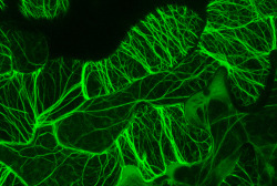
frontal-cortex: Actin microfilaments in epidermal cells of an
frontal-cortex: Microscopy of chicken cells using nano-crystals

mucholderthen: Stress Granules Microphotography by ~lady-alessandra
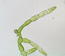
propaedeuticist: protonemata>plant>sporangium: repeat
thisisaadl: It might make you sneeze, but it’s kind of beautiful
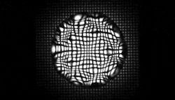
jtotheizzoe: Drop it, Shake It A tiny droplet is vibrated under
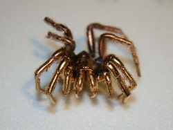
svlc: “A spider coated in gold to prepare it as a specimen
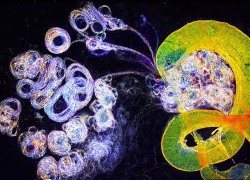
post-mitotic: microscopic bone marrow transplant — hematopoietic
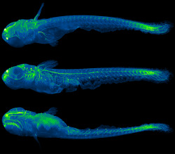
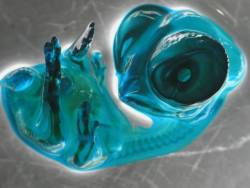
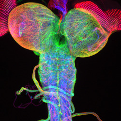
post-mitotic: nervous system of a 5 mm long juvenile medaka
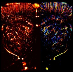
neurosciencestuff: (Image caption: Multiple synapse heads send
sciencenote: Dr. Christian Klämbt and Dr. Imke Schmidt University
txchnologist: Ultrasound Goes Microscopic to Image Living Organs

kqedscience: 6 Amazing Videos From The Olympus Microscopy Competition
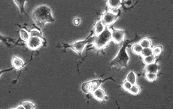
scienceisbeauty: Seeing the Beautiful Intelligence of Microbes
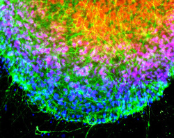
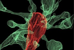
infinity-imagined: Neurons growing in a cell culture These time
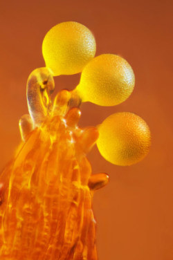
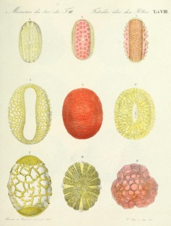
mucholderthen: POLLEN ON THE END OF A FLOWER STAMEN Frederic
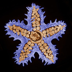
sixpenceee: Starfish imaged using confocal microscopy (10x).(Source)
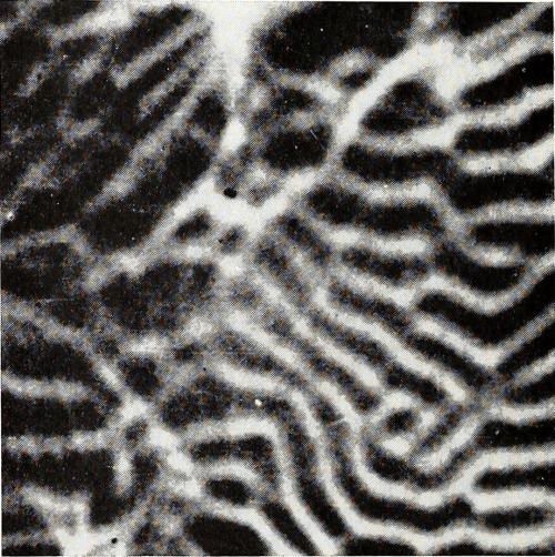
nemfrog: “Electron mirror micrograph of magnetic domain pattern
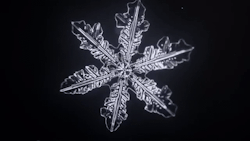
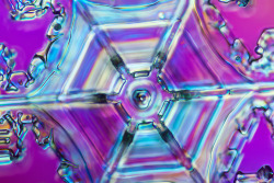
nubbsgalore:snowflake crystallization microscopy (x)

natureofnature: Confocal microscopy of plant tissues

telltaletypist:utopians:utopians:criminal profiling is just astrology

telltaletypist:utopians:utopians:criminal profiling is just astrology
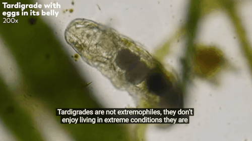
bogleech: witch-niko: Excuse me but since *when* did tardigrades
utopians:utopians:criminal profiling is just astrology for copstaking
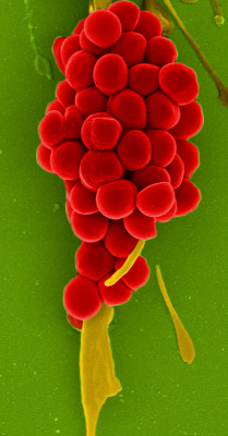
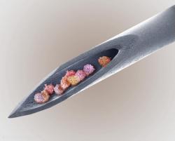
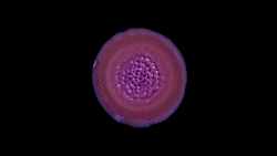
mucholderthen: GRAPES OF WRATHStaphylococcus aureus colonyCourtesy
svlc: “A spider coated in gold to prepare it as a specimen
oxane: Heart of a sectored plate snowflake using DIC microscopy
frontal-cortex: Microscopy of chicken cells using nano-crystals
nothingpersonaluk: Microscopy IV : photo by: © robert gaudette

yeardleysmith: Scanning electron microscopy (SEM) image of a
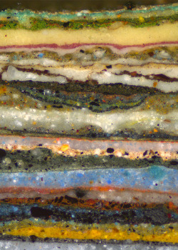
wnycradiolab: A cross-section of wall paints from an 18th century
nubbsgalore: snowflake crystallization microscopy (x)